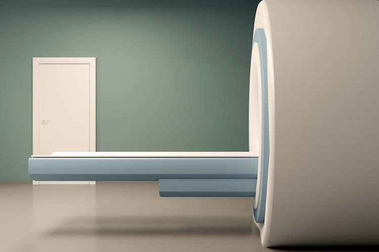
Nuclear medicine is a branch of medicine that uses very small amounts of radioactive materials to diagnose and treat disease. Techniques used in nuclear medicine include:
- Bone scanning;
- PET – Positron Emission Tomography;
- SPECT – Single Photon Emission Computed Tomography;
- Cardiovascular imaging – imaging of the heart and blood vessels.
How does it work?
Nuclear medicine uses radioactive chemicals called radiopharmaceuticals, which are taken into the body, either by injection into a vein, by mouth or by breathing them in. These radiopharmaceuticals continuously give off invisible radiation called gamma rays. Once the radiopharmaceutical has lodged in the relevant part of your body, a special ‘gamma camera’ is used to scan your body and take ‘pictures ‘which record the radiation emitting from your body. These ‘pictures’ are processed by a computer to build up images of the body.
Unlike plain X-rays, and some other diagnostic imaging, which can only show the anatomy and structure of the body, nuclear medicine can show how an organ functions and whether it is working correctly.
Safety and risks
Radiation
The radiopharmaceuticals commonly used in nuclear medicine are safe because:
- They are quickly eliminated from the body.
- They rapidly lose their radioactivity – the materials used have short half-lives (several hours or days).
In addition to being safe, tests using nuclear medicine are also painless.
After having a nuclear medicine test, you will temporarily be weakly radioactive, so you will be asked to avoid close contact with young children for a short time to avoid exposing them to unnecessary radiation. You may be encouraged to drink more fluids to help flush the radiopharmaceutical tracer from your body.
The amount of radiation you are exposed to varies depending on the type of scan.
Pregnancy and breastfeeding
If you are pregnant or breastfeeding, you must tell the doctor or technician. Nuclear medicine studies are often avoided in pregnant women.
Similarly, breastfeeding may need to be avoided for a short period after the test.
Allergic reactions
Allergic reactions to the radiopharmaceutical are very rare and almost always minor.
Bone scans
A common use of nuclear medicine is the bone scan, which can show up fractures (breaks) which may not be visible on normal X-rays. Bone scans are also very useful in detecting arthritis, cancer or infection in the bone.
Blood clots in the lungs
Nuclear scanning can be used to detect blood clots in the lungs (pulmonary embolism).
Heart scans
Blood flow to the heart muscle can be studied through radioactive scanning and this may be very useful in suspected heart attacks and the diagnosis of chest pains, such as angina. Nuclear medicine can also show how well your heart is pumping blood.
PET scans
PET (positron emission tomography) scans are often used to image the heart, brain or cancers in the body. Some cancer cells show up as bright spots on the scan because the cancer cells use more glucose than normal healthy cells. PET scans can show activity in certain areas of the brain, helping to diagnose seizures or tumours.
SPECT scans
Single photon emission computed tomography (SPECT) scans can show blood flow to the heart and activity in the brain. They are used to diagnose dementia, seizures, head injuries, clogged coronary arteries and bone disorders.
Treatment using nuclear medicine
Nuclear medicine is also used for treatment, as well as diagnosis, particularly for cancer. Treatment usually involves swallowing radiopharmaceuticals in capsule or liquid form. Examples of conditions where this form of treatment is used are: thyroid cancer, overactive thyroid gland, some arthritis, certain types of lymphoma, some blood disorders, bone metastases, carcinoid tumours and adrenal gland tumours.

