A biopsy is a procedure in which a small sample of tissue is removed from a particular part of the body so that it can be prepared and examined under a microscope to help diagnose a disease. More advanced tests may also be done on this tissue sample, such as cultures for viruses or other germs. Commonly, biopsies are used to check for cancer, but many other uses exist.
Preparation
Most biopsies don’t require any preparation by the person having the procedure. If a sample is to be taken from the linings of the lungs or the gastrointestinal tract, fasting is often recommended to minimise the risk of vomiting.
The procedure
The exact procedure varies with each biopsy location. A biopsy may be performed through the skin or from inside the body into the region where there might be a problem. Biopsies through the skin may be performed as either ‘open’ or ‘closed’ operations.
An open biopsy means that the skin is cut open and the surgeon parts the tissues to reach the lump or the organ to be examined. A small piece is removed and the incision is then closed. A small lump close under the skin will be biopsied under local anaesthetic but a lump in a more difficult place will involve an operation with a general anaesthetic.
In a closed operation, a special needle is pushed through the skin into the area to be biopsied. The needle then takes up some tissue which is then drawn out. Because the needle is so small the procedure requires only a local anaesthetic and no stitching afterwards.
An internal biopsy means that the doctor can see the organ or part of the body from which a piece of tissue is to be taken without having to perform an open operation. This is done by using a special flexible tube (an endoscope) that has a light and telescopic lens at one end that transmits a picture along the tube’s entire length when inserted into the body. The tube can slide between the body’s organs and allows the doctor to see the internal areas that previously had to be examined by an open operation.
Computerised tomography or ultrasound can also be used to guide certain biopsies of internal organs using special needles that are passed through the skin directly to the organ.
Diagnosis
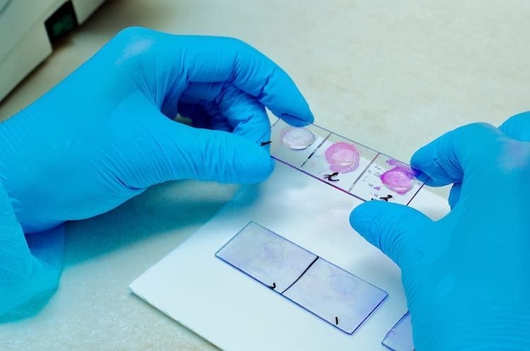
When the tissue is removed, it is usually taken to a pathology laboratory where it is embedded in wax to preserve it. The sample is then thinly sliced, stained a special colour so it can be seen clearly and mounted on a microscope slide. The pathologist looks for normal and abnormal cells. If there are abnormal cells present, it can also be determined what stage the condition has reached.
In some cases, such as during surgery, the tissue is examined soon after it is removed so that the results are available to the surgeon within minutes of the biopsy procedure. Usually, however, the results are available one to two days later.
When is a biopsy necessary?
Biopsies are most often carried out on lumps in the body because it can be difficult to know if a lump is benign (not cancerous) or malignant (cancerous). Biopsy of the liver or kidney may be carried out when the organ is diseased and a more detailed diagnosis is needed before deciding on the appropriate treatment.
What are the risks of a biopsy?
A biopsy is a relatively simple and safe procedure. The spot where the biopsy was taken may be a bit painful when the anaesthetic wears off, but this generally lasts only a day or two. There may be some bleeding and this may cause a discoloration under the skin, but this disappears quite quickly. Bleeding or infection is always a possibility with any type of procedure but this is rare with biopsies. Some organs can be perforated during a biopsy and may require surgery, but this is also very rare.

