What is a PET scan?
A PET (positron emission tomography) scan is a medical imaging technique that provides useful information about the function of organs and tissues within the body. It involves the administration of small amounts of radioactive pharmaceuticals, also known as ‘tracers’. Tracers release invisible energy that can be detected by a special camera to produce 3-dimensional images.
Unlike plain X-rays and other medical imaging techniques, which only provide information on the structure of the body, PET scans can be used to monitor small functional changes over time and can even assess your response to treatment. This is useful in certain diseases, such as cancer, where functional changes may be occurring before any structural changes are evident.
Why is it done?
PET scans can provide detailed information about the function of your organs in one image. Your doctor may order a PET scan to assess blood flow around your body, to differentiate healthy tissue from damaged tissue and to assess the metabolic function of tissues. This is especially useful in assessing cancer, heart disease and brain disease where changes may show up on a PET scan before they appear on other types of imaging (e.g. CT or MRI).
How does it work?
PET scans involve the use of tracers, called radiopharmaceuticals, that are introduced into the body intravenously (by injection into a vein), orally (by mouth) or by inhalation. As tracers break down, they release small positively charged particles known as ‘positrons’. Positrons interact with negatively charged electrons within the body and result in the production of 2 photons. A PET scanner can detect these photon emissions to create images.
There are various types of tracers that can be used depending on the reason the procedure is being done. These include:
- 18-fluorodeoxyglucose (FDG): This is the most commonly used tracer and is derived from glucose, a simple sugar. It is taken up into tissues in different concentrations depending on the metabolic rate of that tissue. It is most commonly used to assess cancer, but can also be used to assess brain diseases and heart diseases. Cancer cells have high metabolic activity and show up as ‘hot spots’ on the PET scan.
- Gallium-68 PSMA: This is most commonly used for prostate cancer staging or restaging.
- Gallium-68 Dotatate: This is most commonly used for neuroendocrine or carcinoid tumour staging or restaging.
Tracers accumulate in areas with high metabolic activity. These areas are known as ‘hot spots’, and appear brighter than the surrounding tissue on the PET scan images. This can provide doctors with vital information about processes that are occurring within the body. For example, healthy heart vessels will take up more tracer than unhealthy areas. So, if certain vessels of the heart are not taking up the tracer as well as other vessels, this may indicate reduced blood flow and raise suspicion of coronary artery disease.
Who should have a PET scan?
A PET scan can be used in the assessment of cancers, brain disease and heart disease.
PET scans for cancer
PET scans can be used in the early detection of cancer, monitoring of disease progression, assessing response to treatment and in detecting metastasis (spread of cancer to other areas of the body).
Radioactive glucose molecules (FDG) are the most common tracers used in the assessment of cancer. Cancer cells appear brighter than normal tissue on the images as they break down glucose at a higher rate. It is also useful in distinguishing between benign (non-cancerous) and malignant (cancerous) tumours.
Several types of cancers can be assessed using PET scans, such as:
- Brain cancer
- Head and neck cancer
- Thyroid cancer
- Colorectal cancer
- Pancreatic cancer
- Breast cancer
- Prostate cancer
- Lymphoma
- Melanoma.
PET scans for heart disease
Doctors can use PET scans to diagnose coronary artery disease (CAD) and to identify areas of reduced blood flow due to narrowed arteries. A PET scan may also identify areas of the heart muscle that have been damaged following a heart attack. The PET scan can distinguish between healthy heart muscle and damaged heart muscle. It can help determine whether there is damaged heart muscle that may be saved if blood flow can be restored. Doctors can use this information to determine what treatment options are best for you, such as coronary artery bypass grafting surgery (CABG) or angioplasty.
PET scans for brain disease
PET scans can be used to assess brain diseases such as cancer, seizures and Alzheimer’s disease. They can provide useful information on blood flow and oxygenation of the brain and highlight areas that aren’t working properly. The brain uses glucose as its main source of fuel, and the PET scan will show up areas of the brain that are using the most radioactive glucose tracer and therefore are the most active.
What happens during a PET scan?
Before the procedure, the nuclear medicine scientist will have a chat with you about the procedure and determine if you have any underlying medical conditions that are important to know about before the procedure, such as diabetes. People with diabetes may need to follow special instructions.
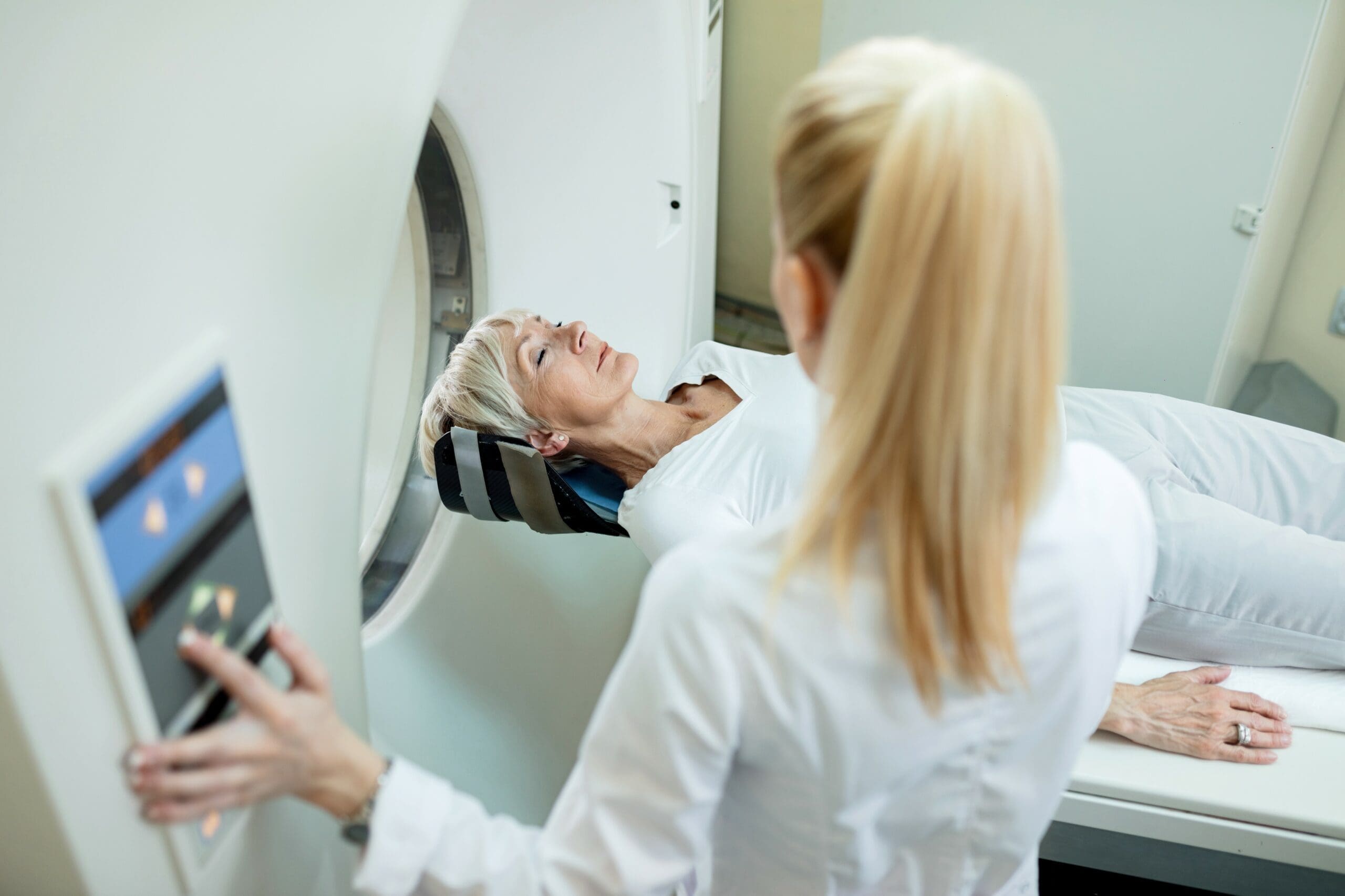
The tracer will either be swallowed, injected or inhaled. You may need to wait 30-60 minutes for the tracer to spread around your body before you undergo the scan. You will be required to wear a hospital gown and will be asked to lie still on a padded bed which is slid into the PET scanner. The scanner will proceed to take numerous images of your body. During this time, the scanner may produce noises such as clicks and buzzing sounds. It is important that you do not move once inside the scanner otherwise the images produced may be blurry.
In most cases, doctors will combine computed tomography (CT) scan with the PET scan, known as ‘hybrid imaging’. The CT scan will be done first and usually takes around 10 minutes to complete. The CT scan can create detailed images of the structure of the body, while the PET scan provides information on the function of the body.
How long does a PET scan take?
The overall time you will spend in the nuclear medicine department or facility may be 2 to 3 hours. But you will only be required to lie on the scanner bed from anywhere between 10 to 40 minutes. Some people may start to feel claustrophobic within the machine. If this happens, tell the doctor or nurse and they may give you medication to help you relax. If you know you are claustrophobic, make sure you discuss this with your doctor before beginning the scan.
What happens after the scan?
The intravenous line will be removed and you should be able to go home.
- Continue with normal daily activities: After you have gone home, you can continue with your normal activities. The tracer should not make you feel sick or drowsy.
- Avoid contact with pregnant or breastfeeding women: You may release a small amount of radiation 6-12 hours after the procedure while the tracer is being removed by your body, so it is advised to avoid contact with pregnant or breastfeeding women.
- Drink plenty of water: FDG is removed from your body via the kidneys and is present in urine. You should drink plenty of water following the procedure to quickly eliminate the tracer from your system. In some cases, the doctor may prescribe a diuretic tablet to help empty your bladder.
How do I prepare for a PET scan?
Your doctor will tell you exactly what you need to do before the PET scan.
Tell your doctor if:
- You have an underlying medical condition, such as diabetes
- You are taking any medications, including over the counter medications, vitamins or supplements
- You have any allergies
- You are pregnant or breast-feeding
- You have had something to eat or drink (other than water) in the last 4-6 hours
- You are claustrophobic.
Generally, you will be required to:
- Avoid strenuous activity for 24 hours before the procedure
- Avoid high carbohydrate and high sugar meals for 24 hours before the procedure. This includes alcohol, bread, rice, cereals, soft drinks and fruit juice
- Fast for 4-6 hours prior to the procedure (including chewing gum or lollies), although plain water is allowed
- Avoid talking for 20 minutes before the procedure
- Remove all jewellery, watches and body piercings. You should not be wearing any metal during the scan, including any on bra straps.
Does a PET scan hurt?
If the tracer is introduced into the body intravenously, you may feel some discomfort from the injection process. The tracer itself is completely painless and you will not feel discomfort during the procedure.
Is a PET scan safe?
A PET scan is generally a safe procedure. Tracers introduced into the body are short-lived and are quickly eliminated. In the time taken for the tracer to be eliminated, you may emit a small amount of radiation. As a result, it is advised you avoid contact with pregnant or breastfeeding women.
In rare circumstances, some people may have a severe allergic reaction to the tracer. It is not possible to predict which people will have an allergic reaction unless they have previously had a reaction to a tracer. All PET scan centres will be prepared to deal with an allergic reaction.
Radiation
The amount of radiation you will be exposed to during a PET scan is equivalent to 3 years of natural background radiation from the environment. However, with repeated scans, the radiation dose can accumulate. Your doctor will ensure the benefits of the procedure outweigh the negative effects so that you are not exposed to unnecessary radiation.
Can you have a PET scan if you are pregnant or breastfeeding?
Please inform your doctor if you are pregnant or breastfeeding. PET scans are generally not recommended for pregnant or breastfeeding mothers.
There is a small risk that your unborn baby will be exposed to radiation during the procedure. Studies are unclear on the exact amount of radiation your baby will be exposed to; however, it is generally low. Your doctor will only advise a PET scan if the benefits to the mother outweigh the harm to the baby. Otherwise, your doctor will consider alternative forms of imaging.
The tracer accumulates in the bladder after the procedure. The bladder is in close proximity to the uterus and is a primary source of radiation to the fetus. If you are pregnant and undergoing a PET scan, ensure you drink plenty of water after the procedure to eliminate the tracer from your bladder. This will minimise radiation exposure to your baby.
If you are breastfeeding, it is recommended to avoid breastfeeding for 6-12 hours after the procedure as the tracer may be present in your breast milk. The exact time frame depends on the procedure and which tracer is being used. To feed your baby during this time, you can express breast milk prior to the procedure or alternatively use formula.
It is also advised to avoid close contact with your baby for 6-12 hours following the procedure.
Do I get the results on the same day?
No, PET scan results are not available straight away. It takes time for the computer to process the information to produce images. Once images have been produced, they will be interpreted by a qualified radiologist who will then send a report to your referring doctor. You will need to make a follow-up appointment with your doctor to receive the results.
What can I expect from the results?
It is difficult to say what you can expect from the results, as it depends on what the doctors are looking for. Generally, doctors will be looking for areas where there is increased uptake of the tracer (‘hot spots’) or areas where the tracer isn’t reaching as well as it should. The doctors may use this information to determine what the next best step should be.
There are certain areas of the body that take up the tracer at a higher rate than other areas, including the brain and the bladder. As a result, these areas normally appear brighter on scan images in all individuals.

