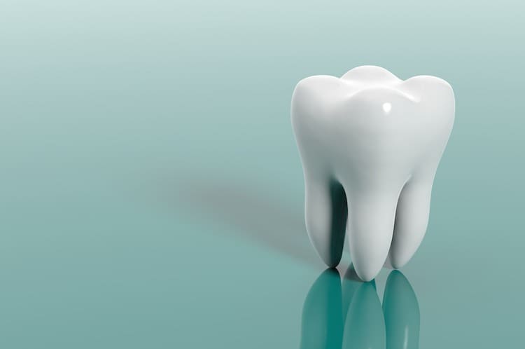
This diagram of a healthy tooth cut in half lengthways shows the layers of the tooth and its internal structure, as well as how the tooth relates to the gum and surrounding jaw bone.
The crown is the part of the tooth that is visible above the gum (gingiva).
The neck is the region of the tooth that is at the gum line, between the root and the crown.
The root is the region of the tooth that is below the gum. Some teeth have only one root, for example, incisors and canine (‘eye’) teeth, whereas molars and premolars have 4 roots per tooth.
The crown
The crown of each tooth has a coating of enamel, which protects the underlying dentine. Enamel is the hardest substance in the human body, harder even than bone. It gains its hardness from tightly packed rows of calcium and phosphorus crystals within a protein matrix structure. Once the enamel has been formed during tooth development, there is little turnover of its minerals during life. Mature enamel is not considered to be a ‘living’ tissue.
Dentine
The major component of the inside of the tooth is dentine. This substance is slightly softer than enamel, with a structure more like bone. It is elastic and compressible in contrast to the brittle nature of enamel. Dentine is sensitive. It contains tiny tubules throughout its structure that connect with the central nerve of the tooth within the pulp. Dentine is a ‘live’ tissue.
Cementum and the periodontal membrane
Below the gum, the dentine of the root is covered with a thin layer of cementum, rather than enamel. Cementum is a hard bone-like substance onto which the periodontal membrane attaches. This membrane bonds the root of the tooth to the bone of the jaw. It contains elastic fibres to allow some movement of the tooth within its bony socket.
The pulp
The pulp forms the central chamber of the tooth. The pulp is made of soft tissue and contains blood vessels to supply nutrients to the tooth, and nerves to enable the tooth to sense heat and cold. It also contains small lymph vessels which carry white blood cells to the tooth to help fight bacteria.
The root canal
The extension of the pulp within the root of the tooth is called the root canal. The root canal connects with the surrounding tissue via the opening at the tip of the root. This is an opening in the cementum through which the tooth’s nerve supply and blood supply enter the pulp from the surrounding tissue.

