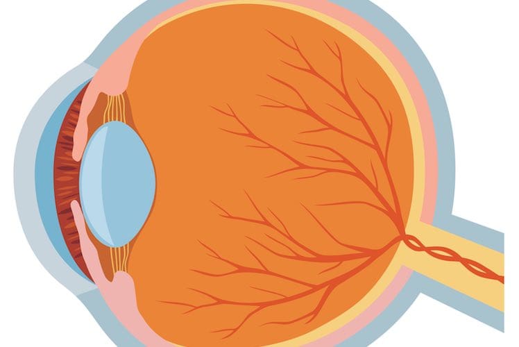
Your eyes sit in sockets within the bones of your skull (known as the orbits), and are surrounded by fat, fibrous tissue and muscles that help protect them from damage. Your eyes are also protected by your eyelids and eyelashes, which block out bright light and help to keep out dust, dirt and other foreign objects.
Tears are produced by the lacrimal gland, located in the orbit just above the outside corner of each eye. Tears are swept across the front of your eyes each time you blink, and drain into ducts at the inner corners of the eyes. Tears not only lubricate the eyes, but also work with your eyelids and eyelashes to protect against dirt and infection.
Structures of the eyeball
The outer layer: the sclera and cornea
The sclera is the tough, fibrous outer layer of the eyeball that forms the whites of your eyes. The front of the sclera is covered by the conjunctiva — a thin, transparent membrane that’s involved in protecting your eyes. The conjunctiva also lines the insides of the eyelids.
The cornea is a dome-shaped structure at the front of the eye. It is transparent, allowing light to enter the eye, and together with the lens helps focus and direct light onto the retina.
The middle layer: the uveal tract (iris, ciliary body and choroid) and lens
The iris is the coloured part of your eye that controls the size of the pupil — the black area in the centre of the iris. When you are in bright light, the iris reduces the pupil size to restrict the amount of light entering the eye; when in dim light or darkness, the iris opens up the pupil to allow more light in.
The iris sits between the anterior chamber and the posterior chamber in the front part of the eye. These chambers contain a watery liquid known as aqueous humour, which is constantly being produced (by the ciliary body) and drained away. Aqueous humour is important for nourishing the lens and cornea.
The lens is a clear, flexible structure that changes shape so that you can focus on objects at varying distances. The lens is connected to the ciliary body (which contains the ciliary muscles) by suspensory ligaments. When the ciliary muscles contract, the normal tension exerted on the lens by the suspensory ligaments is released, and the lens becomes thick and curved, allowing you to see close-up objects clearly. When the ciliary muscles relax, the lens becomes thinner, which is necessary for long-range vision.
The vitreous humour is a jelly-like substance that fills the back portion of the eye behind the lens. As well as helping the eye keep its shape, this clear gel transmits light to the back of the eye.
The choroid, a membrane found between the sclera and the retina, lines the back of the eye. It contains many blood vessels that supply oxygen and nutrients to the retina, and is highly pigmented to help absorb light and prevent scattering.
The inner layer: the retina
The retina lines the inside of the back part of the eye, and is the light-sensitive part of the eye. The retina contains millions of cells known as photoreceptors, and each photoreceptor is linked to a nerve fibre. You have a blind spot, also known as the optic disc, at the back of each eye where all of these nerve fibres converge to form the optic nerve. But this blind spot is usually not noticed, because objects that fall on the blind spot of one eye are seen by the other eye.
Once an image is detected by the photoreceptors, this information is converted into nerve impulses that are sent to the brain via the optic nerve.
The macula is a small area of the retina that contains a high concentration of photoreceptors, and is important for sharp central vision. The middle part of the macula — the fovea — is the most sensitive area, providing the sharpest vision.
Eye movements
The movements of your eyes are controlled by a set of so-called extraocular muscles. These muscles run from the bones of the orbit to the sides of the eyeball. Each eye has 6 muscles to help it move in different directions — the lateral, medial, superior and inferior rectus muscles, and the superior and inferior oblique muscles. People who have a squint (strabismus) may have a problem with one of these muscles or the nerves that control it.
Tears
Tears are usually associated with crying, often as a result of unhappiness or pain. But our eyes make tears continuously. Tears are important for protecting, lubricating and cleaning the eyes.
Tears are secreted by tiny glands (lacrimal glands) situated under the eyelid above the outer corner of each eye.
They flow across the eye, acting as a lubricant. Their spread across the eye is helped by blinking.
Tears leave the eye by draining into 2 tiny canals, the lacrimal ducts (sometimes called tear ducts), situated near the corner of each eye next to the nose. The 2 lacrimal ducts for each eye join together forming the nasolacrimal duct, down which tears flow into the nose.
When things go wrong with this system one of 2 things may happen: the eye may become watery or it may become dry.

